In my 15 years of working in canine physical rehabilitation, iliopsoas strain is the injury I see mismanaged more than any other in the agility population. Dogs come to me weeks after an event, carrying a vague hindlimb lameness that has already been attributed to hip dysplasia, a lumbar disc, or even behavioral reluctance at the start line. The real culprit, a strained iliopsoas complex, is sitting right there waiting to be found. The problem is that most people are not looking for it correctly.
This post is for the rehabilitation professionals, sports medicine veterinarians and experienced handlers who want a clear framework for identifying this injury and a structured protocol for getting these dogs back to full competition safely. I will walk through everything I use in my daily practice at a specialty canine rehabilitation practice, from palpation technique to underwater treadmill progression.
Why This Injury Gets Missed
The iliopsoas does not behave the way most orthopaedic injuries do. There is no joint effusion to flag on radiographs. The lameness can be subtle and intermittent, often worse after rest and improving with a brief warm-up period, which leads handlers to believe the dog is "just stiff." The muscle lies deep in the retroperitoneal space, inaccessible to casual palpation, and the referred pain pattern mimics lumbosacral disease convincingly.
I have reviewed cases where dogs were managed for months under a presumptive diagnosis of early hip dysplasia, including NSAID trials and rest, without resolution. When proper palpation was performed and ultrasound was ordered, the muscle belly and myotendinous junction showed the grade I or II strain that had been there the entire time.
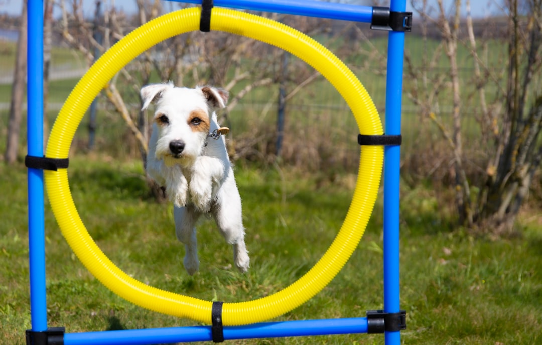
This injury is not rare. In my clinical experience reviewing agility patients, iliopsoas pathology is among the top three soft tissue injuries I encounter in high-drive working dogs. The agility community in particular is vulnerable because the sport demands explosive hip flexion and repeated eccentric deceleration through every single jumping effort.
Anatomy and Mechanism of Injury
The iliopsoas is a functional unit composed of the iliacus and psoas major muscles. In the dog, these muscles originate from the transverse processes of L2 through L6 and the iliac fossa respectively, converge into a common tendon, and insert on the lesser trochanter of the femur. The muscle complex is the primary hip flexor and plays a critical stabilizing role in lumbar spinal extension during the caudal drive phase of gait.
In agility specifically, the mechanism of injury typically falls into one of two patterns. The first is acute eccentric overload, most commonly during weave pole entries, tight serpentine sequences, and collection before a jump. The dog decelerates rapidly while the hip is in a flexed, internally rotated position and the muscle is forced to lengthen under load. The second is chronic repetitive microtrauma, the pattern I see most often, where repeated competition without adequate recovery and structured conditioning allows progressive fiber disruption at the myotendinous junction or the osseous insertion.
The myotendinous junction near the lesser trochanter insertion is the most common site of pathology. Enthesopathy at the lesser trochanter, visible on ultrasound as irregular fiber architecture and peritendinous edema, is a finding I encounter regularly in dogs with multi-season agility careers and insufficient off-season rehabilitation.
Clinical Presentation in Agility Athletes
The presentation varies by grade and chronicity but there are reliable patterns I look for. A dog with an acute grade II strain will typically present with a visible weight-bearing hindlimb lameness that worsens after exercise. The handler often reports the dog "popping out" of the weave poles or refusing jumps with the affected hindlimb as the takeoff leg.
With chronic or low-grade injuries the picture is subtler. The handler notices the dog is slower on course, losing time in collection sequences, or showing what they describe as "ring stress" because the dog appears reluctant to engage. These dogs often have normal gait at a walk and only show dysfunction at trot or on video analysis during sport-specific movements.
My palpation protocol is non-negotiable. With the dog in lateral recumbency on the affected side up, I flex the hip to approximately 90 degrees, apply slight internal rotation, and palpate deeply into the inguinal region following the muscle belly proximally toward the iliac attachment. Guarding, flinching or a pain response on direct pressure at the lesser trochanter constitutes a positive finding. I then perform the stretch test: placing the dog in lateral recumbency with the affected limb down, extending the hip fully while maintaining slight internal rotation. A positive response is resistance to range of motion and muscle guarding before the normal end range of hip extension.
I also assess the contralateral limb. Bilateral involvement is more common than most clinicians expect, particularly in dogs competing through a full season without structured deloading periods.
Differentiating Iliopsoas Strain from Hip Pathology
This differential is where I spend the most diagnostic effort because the clinical overlap with hip dysplasia, coxofemoral osteoarthritis and lumbosacral disease is significant. Getting this wrong costs weeks or months of appropriate treatment.
Hip dysplasia and coxofemoral OA produce pain on Ortolani testing, reduced passive range of hip abduction and palpable crepitus in the joint. Radiographic changes, including subchondral sclerosis, osteophyte formation at the femoral neck and acetabular remodeling, confirm joint pathology. The iliopsoas-strained dog will often have full passive range of hip abduction and a negative Ortolani with pain concentrated specifically on the hip flexion and internal rotation combination and at the lesser trochanter on direct palpation.
Lumbosacral disease deserves careful differentiation. Dogs with lumbosacral stenosis or discospondylitis present with pain on lumbosacral compression, tail head hypersensitivity and often neurological deficits including proprioceptive ataxia and reduced tail tone. The iliopsoas-strained dog typically has a normal neurological examination. The confounding factor is that chronic iliopsoas strain can produce secondary epaxial muscle hypertonicity and paraspinal guarding that mimics lumbosacral pain on superficial examination.
Biceps femoris and gracilis pathology round out the major differentials in the hindlimb. These muscles have distinct palpation sites and functional loss patterns that I assess systematically before committing to an iliopsoas diagnosis.
Imaging and Diagnostic Confirmation
Radiographs are a necessary first step and are ordered by the supervising veterinarian to rule out bony pathology at the lesser trochanter, coxofemoral joint and lumbar spine. I look specifically for any evidence of avulsion at the lesser trochanter, which changes the management course significantly and requires surgical consultation.
Diagnostic ultrasound is my preferred confirmatory tool for iliopsoas pathology. An experienced musculoskeletal ultrasonographer can visualize disrupted fiber architecture, intramuscular hemorrhage in acute cases, areas of fibrosis in chronic presentations and peritendinous fluid accumulation at the insertion. I review ultrasound reports closely because they directly inform where I am in the healing continuum and how aggressively I can progress therapeutic loading.
MRI offers superior soft tissue contrast and is the gold standard for complete characterization of injury grade, particularly when ultrasound findings are equivocal or when concurrent spinal pathology needs to be assessed simultaneously. Access and cost limit its routine use in my patient population but it is the appropriate next step when the clinical picture does not fit a straightforward diagnosis.
My 8-12 Week Return-to-Sport Protocol
I stage return to sport across three phases. The timeline compresses to eight weeks for a confirmed grade I strain with early intervention and extends to twelve or more weeks for grade II injuries, chronic presentations or dogs with significant compensatory muscle dysfunction.
Phase One: Tissue Protection and Pain Management (Weeks 1-3)
The goal of phase one is to reduce inflammatory load, restore pain-free range of motion and prevent deconditioning. All high-impact activity stops. The supervising veterinarian manages the pharmacological component including NSAID therapy and, when indicated, therapeutic injection under ultrasound guidance.
My hands-on work in this phase centers on gentle soft tissue mobilization to the iliopsoas, addressing the compensatory hypertonicity I almost always find in the contralateral quadriceps and ipsilateral hamstring group. I use low-intensity underwater treadmill at water levels chest-height or above to maintain cardiovascular condition and gentle limb loading without axial compression. Walking duration starts at five minutes and progresses based on pain response and gait quality assessed each session.
Therapeutic laser is part of my phase one toolkit for its photobiomodulation effects on tissue healing at the myotendinous junction. I apply it per the parameters established by the supervising veterinarian's prescription.
Phase Two: Progressive Loading and Neuromuscular Re-Education (Weeks 4-7)
Phase two is where the real rehabilitation work happens. Pain at rest should be resolved and palpation should produce only mild discomfort at the insertion site before this phase begins.
I introduce eccentric loading progressions targeting the hip flexor complex through controlled incline walking, cavaletti rail work with deliberate slow cadence and resistance band exercises applied at the stifle during hip extension. The eccentric component is non-negotiable because the mechanism of injury is eccentric overload and the tissue must be conditioned to tolerate that specific stress pattern before return to sport.
Proprioceptive rehabilitation intensifies in this phase. I use balance boards, foam surfaces and controlled lateral weight shifting to address the neuromuscular deficits I consistently find after iliopsoas injury. The muscle's role in lumbopelvic stabilization means that proprioceptive retraining is not optional rehabilitation extra work, it is foundational to injury prevention on return.
Underwater treadmill sessions now include mild incline and increased speed to increase hip flexion demand while maintaining the hydrotherapy benefit of reduced concussive loading. I am watching for compensatory patterns in each session, specifically hip hiking, trunk lateral flexion and asymmetric foot placement, and addressing them before they become ingrained substitution strategies.
Phase Three: Sport-Specific Conditioning and Return Criteria (Weeks 8-12)
Phase three bridges rehabilitation to competition. I work directly with the handler and the supervising DVM to design a progressive return that mirrors the actual demands of agility.
I begin with flat recall work and low jump grid sequences at reduced height, watching the takeoff mechanics on the affected limb closely. Collection and extension sequences are added progressively. Weave poles, the highest-demand obstacle for the iliopsoas, are among the last elements reintroduced and only when the dog is showing symmetric hip flexion mechanics and pain-free palpation at all sites.
My return-to-competition clearance criteria are specific. I need pain-free deep palpation at the lesser trochanter, symmetric passive hip extension range of motion within 10 degrees of the contralateral limb, normal gait at all speeds on video analysis and successful completion of a sport-specific conditioning trial without post-exercise lameness or behavioral reluctance. I will not sign off on return without meeting all four criteria.
Prevention and Long-Term Athlete Management
The dogs I see return to my table with iliopsoas reinjury almost always share one characteristic: they went back to full competition mileage without a structured deloading strategy. Agility handlers are passionate and competitive, which I deeply respect, and the temptation to return to trialing as soon as the dog "looks fine" is real and understandable.
My prevention recommendations for active agility dogs are consistent year to year. Regular structured warm-up including dynamic hip flexor mobilization and controlled trot sets before any agility run. Off-season conditioning that maintains hip flexor eccentric strength through hill work and targeted core stabilization. Annual functional movement screening to catch asymmetries before they become injuries.
I also advocate strongly for periodic musculoskeletal examinations by a veterinarian with sports medicine training, ideally a diplomate of the American College of Veterinary Sports Medicine and Rehabilitation (ACVSMR), as part of the competitive agility dog's annual care plan. Early detection of subclinical iliopsoas tightness and incipient enthesopathy changes the entire trajectory of management.
As a CCRA working under veterinary supervision at a specialty canine rehabilitation practice, my role is to translate the clinical diagnosis into a rehabilitation plan that respects tissue biology and the demands of the sport. Getting the iliopsoas-strained agility dog back to the start line takes patience, precise progressions and honest return-to-sport criteria. The dogs that go through a full, properly staged protocol come back stronger and more durable than before the injury. That outcome is worth every week of the process.